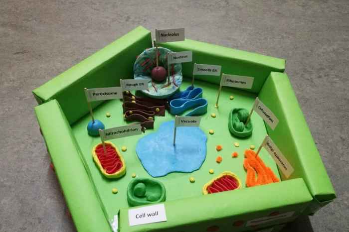
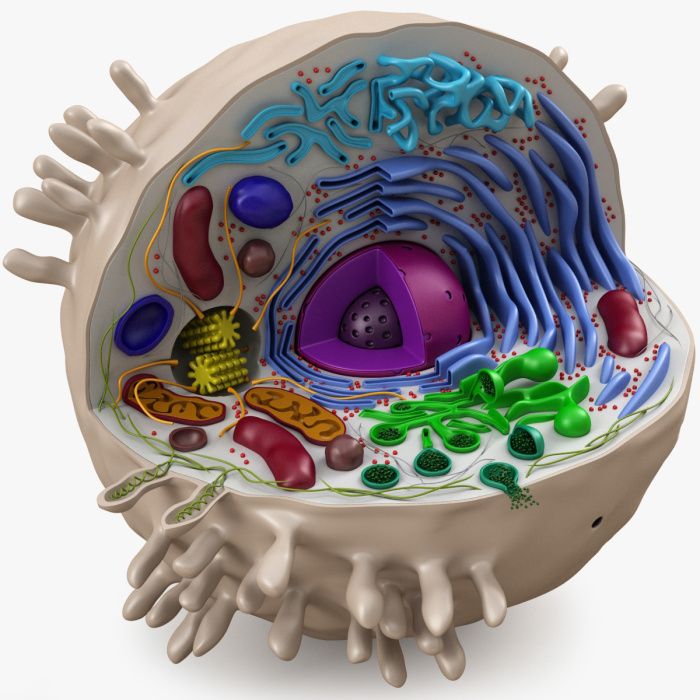
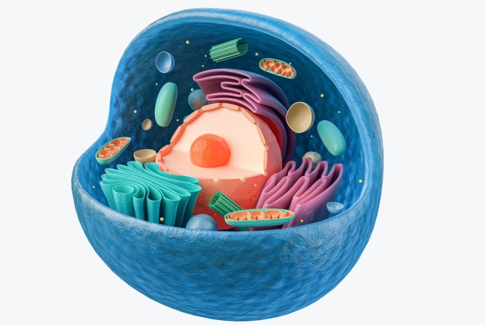
Cells 3D model, a revolutionary approach in biological research, is shaking things up. Forget the flat, boring 2D cell cultures; 3D models mimic the real deal, offering a more realistic environment for studying cells. Think of it as a mini-organ, complete with intricate structures and interactions, all within a tiny space.
This opens up a whole new world of possibilities for understanding how cells behave, how diseases develop, and how new drugs might work.
These 3D models come in various flavors, from spheroids, tiny balls of cells, to organoids, miniature versions of actual organs. Microfluidic chips, like tiny laboratories on a chip, offer another way to create these 3D environments. But how do we make these intricate models?
Well, that’s where the magic of bioengineering comes in.
Introduction to 3D Cell Models
In the realm of biological research, understanding the intricate workings of cells is paramount. Traditional 2D cell culture methods, while valuable, often fail to capture the complexity and dynamic nature of cells within a living organism. Enter 3D cell models, a revolutionary approach that mimics the three-dimensional environment of cells in vivo, providing a more physiologically relevant platform for studying cellular behavior.
Significance of 3D Cell Models in Biological Research
3D cell models have emerged as indispensable tools in biological research, offering a paradigm shift in our understanding of cellular processes. By recreating the three-dimensional architecture of tissues, 3D models provide a more realistic representation of cell-cell interactions, signaling pathways, and responses to stimuli, ultimately leading to more accurate and reliable experimental results.
Advantages of 3D Cell Models Over Traditional 2D Cell Culture
- Enhanced Physiological Relevance:3D models more closely resemble the in vivo environment, providing a more accurate representation of cellular behavior and responses.
- Improved Cell-Cell Interactions:3D models facilitate complex cell-cell interactions, mimicking the intricate communication networks found in tissues.
- Enhanced Matrix Interactions:Cells in 3D models interact with extracellular matrix components, replicating the structural support and signaling cues present in tissues.
- Improved Drug Discovery and Development:3D models provide a more realistic platform for drug screening and evaluation, leading to more accurate predictions of efficacy and toxicity.
Types of 3D Cell Models
- Spheroids:Spheroids are three-dimensional aggregates of cells that self-assemble into spherical structures. They are relatively simple to create and provide a basic model for studying cell-cell interactions and tumor growth.
- Organoids:Organoids are more complex 3D models that mimic the structure and function of specific organs. They are generated from stem cells or primary cells and can be used to study organ development, disease mechanisms, and drug responses.
- Microfluidic Chips:Microfluidic chips are devices that create miniature 3D environments for cell culture. They allow for precise control of cell microenvironment, including nutrient delivery, waste removal, and mechanical stimulation.
Methods for Creating 3D Cell Models

The creation of 3D cell models involves various techniques, each with its own advantages and limitations. These methods can be broadly categorized into scaffold-based and scaffold-free approaches.
Scaffold-Based Methods
Scaffold-based methods utilize biocompatible materials to provide structural support for cells, allowing them to form three-dimensional structures. These scaffolds serve as templates for cell growth and organization, mimicking the extracellular matrix of tissues.
- Hydrogels:Hydrogels are cross-linked polymer networks that can swell and absorb water, creating a three-dimensional environment for cell growth. They are biocompatible, biodegradable, and can be tailored to mimic the mechanical properties of different tissues.
- Collagen:Collagen is a naturally occurring protein that forms the major structural component of the extracellular matrix. It can be used to create scaffolds that promote cell adhesion, proliferation, and differentiation.
Scaffold-Free Methods, Cells 3d model

Scaffold-free methods rely on the self-assembly of cells or bioprinting techniques to create three-dimensional structures. These methods eliminate the need for external scaffolding materials, offering a more natural approach to 3D cell culture.
- Spheroid Formation:Spheroids can be generated through self-assembly, where cells aggregate and form spherical structures. This process can be facilitated by using specific culture conditions or microfluidic devices.
- Bioprinting:Bioprinting is a technique that uses 3D printing technology to deposit cells and biomaterials in a layer-by-layer fashion, creating complex 3D structures. Bioprinting allows for precise control over cell placement and tissue architecture.
Applications of 3D Cell Models
3D cell models have revolutionized various fields, offering unprecedented opportunities for advancing scientific research and improving human health.
Drug Discovery and Development
3D cell models have become invaluable tools in drug discovery and development, providing a more realistic platform for screening drug candidates and evaluating their efficacy and toxicity. By mimicking the in vivo environment, 3D models allow researchers to assess drug effects on cells in a more physiologically relevant context, leading to more accurate predictions of drug behavior in humans.
- Drug Screening:3D cell models can be used to screen large libraries of drug candidates, identifying those with the most promising therapeutic potential. This process can significantly reduce the time and cost associated with drug discovery.
- Toxicity Testing:3D models provide a more accurate assessment of drug toxicity, allowing researchers to identify potential side effects and optimize drug formulations to minimize adverse effects.
Disease Modeling
3D cell models have proven to be highly effective in modeling human diseases, providing insights into disease mechanisms and facilitating the development of personalized therapies. By recapitulating the cellular and molecular features of specific diseases, 3D models offer a powerful tool for studying disease progression, identifying potential drug targets, and evaluating the efficacy of new treatments.
- Cancer Research:3D models have been widely used to study cancer cell growth, invasion, and metastasis, providing insights into the complex biology of cancer and identifying potential therapeutic targets.
- Neurological Disorders:3D models of brain tissues have been instrumental in studying neurodegenerative diseases, such as Alzheimer’s and Parkinson’s, and developing novel therapies.
Tissue Engineering

3D cell models hold immense promise for tissue engineering, the field of creating functional tissues for transplantation or regenerative medicine. By using biocompatible materials and specialized cell types, researchers are developing 3D models that can be used to regenerate damaged tissues and organs, offering hope for patients suffering from various debilitating conditions.
- Skin Regeneration:3D models of skin tissues are being developed for burn victims and patients with chronic wounds, providing a source of viable cells for transplantation.
- Cartilage Regeneration:3D models of cartilage tissues are being explored for the treatment of osteoarthritis and other cartilage-related conditions.
Challenges and Future Directions
While 3D cell models offer tremendous potential, they also face several challenges that need to be addressed to fully realize their promise.
Technical Challenges
- Scaling Up Production:One of the major challenges is scaling up the production of 3D cell models to meet the growing demand for research and clinical applications. Developing efficient and cost-effective methods for large-scale production is crucial.
- Standardization and Reproducibility:Ensuring the standardization and reproducibility of 3D cell models is essential for generating reliable and comparable data across different laboratories. Establishing standardized protocols and quality control measures is crucial for advancing the field.
Future Directions
Despite the challenges, the future of 3D cell modeling is bright, with ongoing advancements and emerging technologies poised to revolutionize the field.
- Personalized Medicine:3D cell models are expected to play a significant role in personalized medicine, allowing for the development of tailored therapies based on an individual’s genetic makeup and disease profile.
- Organ-on-a-Chip:Organ-on-a-chip technology, which uses microfluidic devices to create functional models of human organs, is a rapidly evolving field with immense potential for drug discovery, disease modeling, and personalized medicine.
- Bioprinting Advancements:Advancements in bioprinting technology, such as the development of bioinks and biocompatible materials, are paving the way for more complex and functional 3D cell models.
Final Summary
3D cell models are not just a cool new trend; they are changing the game. From drug discovery and disease modeling to tissue engineering, these tiny worlds are unlocking secrets and paving the way for new treatments. While challenges remain, the future of biological research is looking pretty 3D.
FAQ Section: Cells 3d Model
What are the limitations of 3D cell models?
While 3D cell models are incredibly powerful, they are not perfect. One limitation is the difficulty in replicating the complex microenvironment of tissues, including factors like blood vessels and immune cells. Additionally, scaling up the production of 3D models for research and clinical applications can be challenging.
How are 3D cell models used in drug discovery?
3D cell models allow researchers to test drug candidates in a more realistic environment, helping them assess efficacy and toxicity. They can also be used to identify potential drug targets and develop personalized therapies.